Full range of orthopedic services from diagnosis to rehabilitation. We provide assistance with injuries and diseases of the joints, as well as sports injuries.
Біль у плечі та руці: чим зумовлений та як лікується
Лікування болю у плечі підбирають після визначення причини симптомів. Однією з найпоширеніших причин болю є перевантаження ротаторної манжети.
#shoulder
Внутрішньосуглобові ін'єкції: призначення та ризики
Внутрішньосуглобові ін'єкції є поширеним методом допомоги при болю у суглобах. Вони можуть швидко полегшити стан, відновивши рухливість і працездатність.
#knee
#shoulder
#thigh
Хвороба Бехтерева: що це, симптоми та діагностика
Хвороба Бехтерева, або анкілозуючий спондиліт — це хронічне запальне захворювання хребта та крижово-клубових суглобів, яке поступово обмежує рухливість і змінює якість життя.
#back
Остеоартроз суглобів: симптоми, причини та лікування
Остеоартроз розвивається тоді, коли суглоб роками отримує більше механічного навантаження, ніж здатен витримати. Суглобова щілина звужується. Кісткова тканина реагує ущільненням і кістковими розростаннями.
#hand
#knee
#elbow
#shoulder
#thigh
Ревматоїдний артрит: симптоми та ревматоїдний фактор
Ревматоїдний артрит — системне аутоімунне запалення, яке вражає суглоби, сухожильні оболонки та інші тканини організму.
#hand
Хвороба Де Кервена: симптоми та методи лікування
Хвороба Де Кервена або тендовагініт — це патологія першого компартменту розгиначів, при якій сухожилля великого пальця проходять крізь звужену ділянку й починають ковзати з болем.
#hand
Hygroma: causes and effective treatment
A hygroma is a superficial, bony formation near a joint or tendon canal that most often appears on the hand, wrist, or fingers.
#hand
Stenosing ligamentitis (clicking finger)
Stenosing ligamentitis disrupts the normal sliding of the tendon of the finger and gradually complicates the usual movements of the hand. The person first feels stiffness, then painful clicks appear, and in the later stages the finger is fixed in a bent position.
#hand
Why Does a Finger Hurt: Main Causes and Treatment
Pain in the finger appears after injury, overload, inflammation or compression of the nerve. The symptom itself does not indicate one specific problem, because the source of discomfort can be a joint, tendons, ligaments, nerve or surrounding soft tissues.
#hand
Joint contracture: causes, symptoms, types and treatments
Joint contracture is a steady decrease in the amplitude of movements due to changes in soft tissues and other structures around them. This condition interferes with the performance of the usual actions and can significantly impair the quality of life.
#hand
Can you run with osteoarthritis?
When running with arthrosis is possible, in which cases it is strictly not recommended, and what alternatives should be considered in order to maintain movement without harm to the joints - says Vasily Mellen, orthopedic surgeon and traumatologist of Mashtaler Ortho&Trauma Clinic.
#knee
What are compression stockings and who needs them?
This article is about what compression stockings are, why they are important in orthopedics, and how we apply them in an integrated approach to recovery in Mashtaler Ortho&Trauma Clinic.
#foot
#knee
Rehabilitation and life after hip endoprosthetics
How life changes after hip replacement, what to expect in the first weeks, what to strive for next and how to make recovery not only effective, but also natural.
#thigh
Bikini access during hip endoprosthetics
Bikini approach is a relatively new, minimally invasive method that is distinguished by its delicacy, less trauma and expressive aesthetics.
#thigh
The First Meniscus Transplant in the Western Region
This is an important achievement, because such an operation was carried out only for the eighth time in Ukraine.
No items found.
Endoprosthetics of the shoulder joint
Shoulder joint replacement surgery is performed to relieve pain and other symptoms that arise due to damage to the shoulder joint. The operation involves removing the affected areas of the bone and replacing them with parts — special implants made of metal or plastic.
#shoulder
Arthroscopy of the ankle
Arthroscopy of the ankle joint is a surgical procedure that allows the doctor to see all components of the joint, with minimal trauma to the surrounding tissues and a low risk of complications.
No items found.
Halus-valgus
Halus valgus or valgus deformity of the big toe is a pathological condition in which the thumb shifts to the side and looks like a large “bulging” growth. It is a condition that can significantly restrict movement and cause pain when walking.
#foot
PCV rupture: causes, mechanisms, symptoms — everything you need to know
Ligaments are strong fibers that connect bones together. The anterior cruciate ligament is one of two ligaments that cross at the center of the knee and connect the femur to the tibia. The anterior cruciate ligament is the main stabilizer of the knee joint. Injuries leading to its rupture provoke further instability of the knee.
#knee
High-Intensity Laser Therapy
High-intensity laser therapy is a powerful method of overcoming pain. På (biostimulation a fotomechanical effect), the laser accelerates the healing and regeneration processes. High-intensity laser technology is based on the principle of low-level laser therapy.
#hand
#knee
#thigh
#elbow
#shoulder
Super-inductive magnetotherapy
Superinductive magnetotherapy (also called High-Intensity Magnetotherapy) is a modern, effective and safe method of treating pain without direct intervention in the human body. This is possible due to the influence on the neuromuscular tissue of the magnetic field, which creates a special superinductive apparatus. Already after the first procedure, there is a noticeable reduction in pain.
#hand
#knee
#thigh
#elbow
#shoulder
BMAC
Nós,. BMAC regenerativna terapija je upravljanje, ki se.
#hand
#knee
#thigh
#elbow
#shoulder
Arthroscopy of the shoulder joint
Arthroscopy of the shoulder joint is a surgical procedure that allows the doctor to see all components of the joint, with minimal trauma to the surrounding tissues and a low risk of complications.
A hygroma is a superficial, bony formation near a joint or tendon canal that most often appears on the hand, wrist, or fingers. It does not belong to malignant tumors, but can cause discomfort, interfere with movements and create doubts if a person notices a new swelling under the skin.
In this article, we will analyze how hygroma is formed, what factors contribute to its appearance, what symptoms are characteristic of this condition and when a specialist examination is needed. We will also explain how the diagnosis, observation, removal and recovery are carried out after the intervention.
What is a hygroma?
Hygroma is a cystic formation filled with a thick jelly-like fluid associated with a joint capsule or tendon sheath. The most typical areas of its development are the back or surface of the wrist, the base of the finger and the terminal joint of the finger.
The question of what it is, a hygroma, most often arises when a person feels a rounded elastic formation in the joint area, which changes in size or becomes more noticeable after a load. Not only shape and size are important for clinical evaluation, but also the relationship with movements, pain, sensitivity and function of the hand.
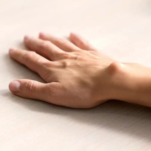
Hygroma on the hand in the form of swelling on the hand
The mechanism of formation of growths
The cyst originates from the tissues surrounding the joint, tendon sheath or ligament structures, and has connections with deeper tissues through the leg or canal. Inside contains a viscous content similar in properties to the fluid that lubricates the joints.
In international literature, this formation is also called “ganglion” or “ganglion cyst”. Its appearance is not due to infection and not to oncological growth, but to local changes in the capsule, tendon canal and synovial fluid.
The size of the cyst is not stable. Against the background of active load on the joint, it sometimes increases, and at rest it becomes smaller or even temporarily almost imperceptible.
How does hygroma differ from other tumor-like formations?
Hygroma usually has smooth contours, lies under the skin, is associated with a joint or tendon and often changes size over time. Unlike hard bony growths, it is not part of the bone, and unlike the epidermoid cyst, it usually does not have a characteristic central opening on the skin.
A growth on a finger is not necessarily one specific formation. Under such a complaint may be hidden:
hygroma;
mucous cyst of the end joint;
fibrokeratoma;
lipoma;
other soft tissue formations.
Therefore, the final conclusion is made after examination and, if necessary, ultrasound. For additional examination, masses that grow rapidly, lie deep, have a diameter of 5 cm or more, do not shift well relative to deep tissues or appear suddenly for no apparent reason are especially important. Such signs are not typical of a simple superficial cyst and require extended evaluation.
Causes of hygroma
An exact universal trigger for each patient has not been established. Possible factors include repeated loads, injuries, microtraumas and associated degenerative changes in the joint or inflammatory changes as factors leading to the formation of such a formation.
Joint overload and repetitive movements
The most famous example is the constant stress on the hand in athletes who work a lot with wrist extension and support. Hygromas are common among gymnasts who repeatedly load the hand and wrist joint.
In everyday life, a similar situation occurs when working with tools, prolonged force grip, repeated squeezing of objects or monotonous small movements. It is these actions that maintain local irritation in the area of the joint capsule or tendon canal.
Injuries and microtraumas
A single injury to the hand is not a prerequisite for the appearance of a cyst, but it can precede its formation. Ganglion cysts can occur against the background of damage to the joint, ligamentous apparatus or tendon structures.
Microtraumas work differently. They do not give a single bright episode of pain, but for a long time maintain changes in the tissues, which eventually form a synovial cyst.
Inflammatory diseases of joints and tendons
Mucous cysts near the end joint of the finger are often associated with early manifestations of osteoarthritis of this joint. This is a separate clinical option that can be accompanied by a furrow or deformation of the nail due to pressure.
Inflammatory changes in tendons and periarticular tissues are also included in the list of causes. In such situations, the formation is formed not in isolation, but against the background of an already existing irritation or degenerative process in the area of the tendon or joint capsule.
Is there a hereditary predisposition?
For ordinary hygromas of the hand, there is no convincingly confirmed hereditary mechanism. The main emphasis in clinical sources is not on genetics, but on local load, anatomical area, and associated joint changes.
A separate nuance concerns the mucous cysts of the end joint of the finger. They are more common in women aged 40 to 70 years and are associated with degenerative changes in the distal interphalangeal joint.
Symptoms indicating hygroma
The most noticeable symptom is a visible or palpable rounded formation near the joint or tendon. Additional complaints depend on the location, size, pressure on surrounding tissues and whether the cyst affects nerves or blood vessels.
External manifestations
The hump on the finger needs to be examined, since not every surface formation is a hygroma. A ganglion cyst is more characterized by a smooth surface, elastic consistency, location near the joint and the absence of signs of inflammatory infection, if the skin over the formation has not reddened and become hot.
Signs that the doctor assesses at the reception:
rounded or oval swelling under the skin;
location in the area of the wrist, base of the finger or the end interphalangeal joint;
resizing after loading on the brush;
smooth contours without pronounced unevenness of the skin;
sometimes transparency when illuminated, if we are talking about a superficial cyst.
As a rule, a small cyst without compression of the nerve structures does not hurt at rest. However, the absence of pain does not cancel the need for examination if the formation grows, becomes denser or changes shape.
Pain and restriction of movements
Pain syndrome does not appear in all patients. If the cyst presses on the nerve, prevents the tendon from sliding, or is located where the joint is actively working, it causes pain, aching discomfort after movement, tingling, weakness in the grip and stiffness.
Symptoms are especially noticeable with hygroma at the base of the finger or near the tendon canal. In such a zone, even a small formation interferes with the compression of objects and makes painful household actions associated with a power grip.
When hygroma can cause complications
Serious complications do not occur in every case, but a clear list of problems is well known to doctors. It includes:
nerve compression with pain or tingling;
decrease in hand strength; limitation of movements;
deformation of the nail with the mucous cyst of the finger;
leakage of contents through thinned skin over the formation on the end joint of the finger.
Separately, redness, increased skin temperature, sharp pain or a hard fixed node are alarming. For such signs, it is necessary to consult a doctor without delay, since a calm picture is no longer typical for an ordinary superficial cyst.
How is the diagnosis done?
In most cases, diagnosis begins with an examination. The doctor clarifies how long ago the formation appeared, whether it changes in size, whether there is pain, numbness, restriction of movements and previous injuries to the hand.
Orthopedist Review
During the examination, localization, shape, mobility, density, connection with joint movements, skin condition and the presence of neurological symptoms are assessed. For the selection of tactics, it is also important to understand whether the formation lies superficially, whether it is adjacent to important structures, in particular nerves and blood vessels.
Signs in which the patient should make an appointment faster:
formation increases for a short time;
pain, tingling or weakness of the hand appeared;
the node has become solid and almost does not shift;
the skin over it reddened or became hot;
the tumor-like mass is located deep or exceeds about 5 cm.
Ultrasound of soft tissues
Ultrasound of soft tissues is used when it is necessary to confirm the cystic nature of the formation, assess its size, depth, boundaries and relationship with surrounding structures. Ultrasonography helps to assess whether the mass is cystic or solid, and also shows the vessels associated with it.
This is especially useful if outwardly the picture is not quite typical or the formation lies next to the vascular-nerve bundle. For palmar cysts of the wrist and formations near the finger, such information affects both the diagnosis and the choice of further tactics.
Methods of treatment of hygroma
Tactics depend on localization, symptoms, rate of change and impact on the function of the hand. If the formation is small, does not cause pain and does not restrict movement, observation is allowed. If the cyst interferes with the work of the hand or recurs after a puncture, surgical intervention is considered.
Observation without surgery
For asymptomatic or minimally painful cysts, observation is considered a reasonable approach. World health organizations agree that a significant proportion of such formations do not require immediate removal and can decrease or disappear on their own.
But conservative treatment does not equal inaction. It includes reducing the load on the hand, temporary immobilization with an orthosis or bandage for pain syndrome, symptomatic anesthesia and control in dynamics.
Puncture with aspiration also belongs to non-surgical options, but its effectiveness is limited. After aspiration, relapse occurs often, and with palmar cysts of the wrist, the method is not always safe due to the proximity of nerves and blood vessels.
It is impossible to pierce or crush the cyst yourself. Trying to pierce it at home increases the risk of skin damage and infection.
Surgical removal
Surgery is considered when the formation causes persistent pain, interferes with function, returns after puncture, or is constantly disturbing due to localization and symptoms. The essence of the intervention is to remove not only the cyst itself, but also its leg or the area of the capsule from which it emerges.
The intervention is usually performed under anesthesia as an outpatient or day procedure. For cysts around the finger, surgery is often performed under local anesthesia, while for wrist formations, regional or, in some cases, general anesthesia is more often used due to a deeper connection with the joint.
No method eliminates the likelihood of relapse. With posterior carpal and dorsal cysts, it is 10%, with palmar carpal cysts — 30%. After aspiration, relapses occur more often — more than 30— 40%.
Recovery after hygroma removal
The postoperative period depends on the localization of the cyst, the amount of intervention and the individual reaction of the tissues. Even when the removal is performed technically without complications, the patient needs some time to heal the skin, relieve swelling and return a comfortable grip.
Healing period
Returning to the usual actions after excision of the cyst often takes from two to six weeks. Dressings and suture removal or wound control often occur on days 10—14, and scar sensitivity and swelling sometimes persist longer.
After surgery, local pain, swelling, a temporary decrease in grip strength and sensitivity of the scar are possible. These are typical phenomena and that is why the doctor explains to the patient in advance the expected course of recovery and the limits of the load on the arm.
Limitation of physical activity
In the first days, the hand is usually protected from the force load, and the bandage is kept dry in accordance with the recommendations of the clinic. Temporary fixation for 1-2 weeks is sometimes applied to the wrist, while a complete return to sports or heavy manual work is postponed until the surgeon examines.
If the work involves tools, lifting loads or a long power grip, the return takes longer than in the office mode. Physically hard work is postponed for several weeks until pain and swelling decrease.
How to reduce the risk of recurrence
Relapse is associated not only with the fact of the operation itself, but also with the type of cyst, its location and the condition of the surrounding joint. That is why the doctor assesses whether there is arthrosis, tendon irritation, pressure on the nerve or zones of constant mechanical friction nearby.
After healing, it is advisable:
dose repeated brush movements and reduce prolonged force grip;
do not return to heavy loads earlier than the doctor allowed;
control the condition of the scar, swelling and sensitivity of the hand after surgery;
re-consult a specialist if the formation has reappeared or the nature of the pain has changed.
Frequently Asked Questions About Hygroms
Can hygroma disappear on its own without treatment?
Yes, this situation is described in several authoritative sources. A ganglion cyst can disappear without treatment within a few months. Some formations do not require immediate intervention and can decrease spontaneously.
However, conservative treatment is not suitable for everyone. If the mass is painful, grows, disturbs movements or raises doubts about the diagnosis, it is better to consult a doctor immediately.
Is hygroma removal painful and which method is most effective?
During the operation, anesthesia should remove the pain at the very moment of the intervention. Discomfort is expected after the procedure, when anesthesia passes, but for this, standard postoperative anesthesia and a gentle load regimen are prescribed.
If we compare other methods with surgery, then observation is appropriate for asymptomatic cysts. Aspiration is not always suitable and has a higher frequency of reappearance. Surgical removal is considered as a more radical option for recurrent formations, since it makes it possible to remove both the cyst and its base.
What is the probability of recurrence of hygroma after treatment?
Reappearance is possible after any tactic, but its frequency varies depending on the localization and method of treatment. For part of the posterior cysts, it is about 10% after surgery, for the palmar carpal — about 30%, and after aspiration, relapses generally occur much more often.
If the formation has returned, you should not decide for yourself that this is a “safe cyst”. Repeated examination is needed in order to assess the source of symptoms, the condition of the joint and the advisability of further observation, puncture or surgery.
.png)