.png)

EN
Grazie! Your submission has been received!
Oops! Something went wrong while submitting the form.

Skript

EN
Nothing found
Full range of orthopedic services from diagnosis to rehabilitation. We provide assistance with injuries and diseases of the joints, as well as sports injuries.
Біль у плечі та руці: чим зумовлений та як лікується
Лікування болю у плечі підбирають після визначення причини симптомів. Однією з найпоширеніших причин болю є перевантаження ротаторної манжети.
Внутрішньосуглобові ін'єкції: призначення та ризики
Внутрішньосуглобові ін'єкції є поширеним методом допомоги при болю у суглобах. Вони можуть швидко полегшити стан, відновивши рухливість і працездатність.
Хвороба Бехтерева: що це, симптоми та діагностика
Хвороба Бехтерева, або анкілозуючий спондиліт — це хронічне запальне захворювання хребта та крижово-клубових суглобів, яке поступово обмежує рухливість і змінює якість життя.
Остеоартроз суглобів: симптоми, причини та лікування
Остеоартроз розвивається тоді, коли суглоб роками отримує більше механічного навантаження, ніж здатен витримати. Суглобова щілина звужується. Кісткова тканина реагує ущільненням і кістковими розростаннями.
Ревматоїдний артрит: симптоми та ревматоїдний фактор
Ревматоїдний артрит — системне аутоімунне запалення, яке вражає суглоби, сухожильні оболонки та інші тканини організму.
Хвороба Де Кервена: симптоми та методи лікування
Хвороба Де Кервена або тендовагініт — це патологія першого компартменту розгиначів, при якій сухожилля великого пальця проходять крізь звужену ділянку й починають ковзати з болем.
Hygroma: causes and effective treatment
A hygroma is a superficial, bony formation near a joint or tendon canal that most often appears on the hand, wrist, or fingers.
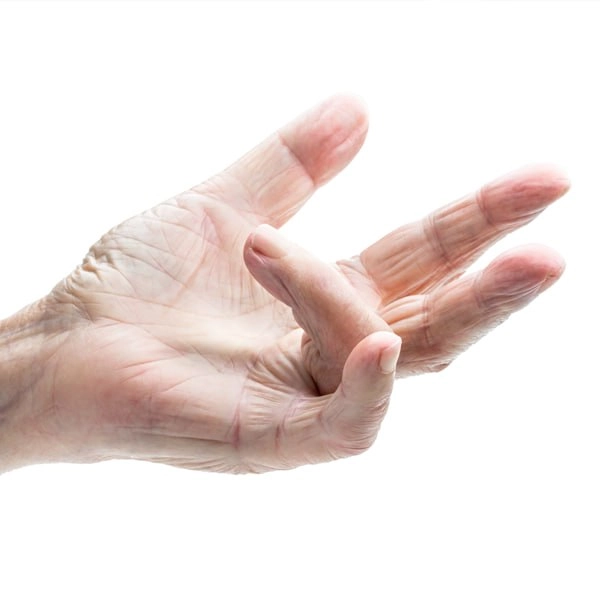
Stenosing ligamentitis (clicking finger)
Stenosing ligamentitis disrupts the normal sliding of the tendon of the finger and gradually complicates the usual movements of the hand. The person first feels stiffness, then painful clicks appear, and in the later stages the finger is fixed in a bent position.
Why Does a Finger Hurt: Main Causes and Treatment
Pain in the finger appears after injury, overload, inflammation or compression of the nerve. The symptom itself does not indicate one specific problem, because the source of discomfort can be a joint, tendons, ligaments, nerve or surrounding soft tissues.
Joint contracture: causes, symptoms, types and treatments
Joint contracture is a steady decrease in the amplitude of movements due to changes in soft tissues and other structures around them. This condition interferes with the performance of the usual actions and can significantly impair the quality of life.
Can you run with osteoarthritis?
When running with arthrosis is possible, in which cases it is strictly not recommended, and what alternatives should be considered in order to maintain movement without harm to the joints - says Vasily Mellen, orthopedic surgeon and traumatologist of Mashtaler Ortho&Trauma Clinic.
What are compression stockings and who needs them?
This article is about what compression stockings are, why they are important in orthopedics, and how we apply them in an integrated approach to recovery in Mashtaler Ortho&Trauma Clinic.
Rehabilitation and life after hip endoprosthetics
How life changes after hip replacement, what to expect in the first weeks, what to strive for next and how to make recovery not only effective, but also natural.
Bikini access during hip endoprosthetics
Bikini approach is a relatively new, minimally invasive method that is distinguished by its delicacy, less trauma and expressive aesthetics.
The First Meniscus Transplant in the Western Region
This is an important achievement, because such an operation was carried out only for the eighth time in Ukraine.
Endoprosthetics of the shoulder joint
Shoulder joint replacement surgery is performed to relieve pain and other symptoms that arise due to damage to the shoulder joint. The operation involves removing the affected areas of the bone and replacing them with parts — special implants made of metal or plastic.
Arthroscopy of the ankle
Arthroscopy of the ankle joint is a surgical procedure that allows the doctor to see all components of the joint, with minimal trauma to the surrounding tissues and a low risk of complications.
Halus-valgus
Halus valgus or valgus deformity of the big toe is a pathological condition in which the thumb shifts to the side and looks like a large “bulging” growth. It is a condition that can significantly restrict movement and cause pain when walking.
PCV rupture: causes, mechanisms, symptoms — everything you need to know
Ligaments are strong fibers that connect bones together. The anterior cruciate ligament is one of two ligaments that cross at the center of the knee and connect the femur to the tibia. The anterior cruciate ligament is the main stabilizer of the knee joint. Injuries leading to its rupture provoke further instability of the knee.
High-Intensity Laser Therapy
High-intensity laser therapy is a powerful method of overcoming pain. På (biostimulation a fotomechanical effect), the laser accelerates the healing and regeneration processes. High-intensity laser technology is based on the principle of low-level laser therapy.
Super-inductive magnetotherapy
Superinductive magnetotherapy (also called High-Intensity Magnetotherapy) is a modern, effective and safe method of treating pain without direct intervention in the human body. This is possible due to the influence on the neuromuscular tissue of the magnetic field, which creates a special superinductive apparatus. Already after the first procedure, there is a noticeable reduction in pain.
BMAC
Nós,. BMAC regenerativna terapija je upravljanje, ki se.
Arthroscopy of the shoulder joint
Arthroscopy of the shoulder joint is a surgical procedure that allows the doctor to see all components of the joint, with minimal trauma to the surrounding tissues and a low risk of complications.